Recognizing Early Signs of Shell Rot in Aquatic and Terrestrial Chelonians
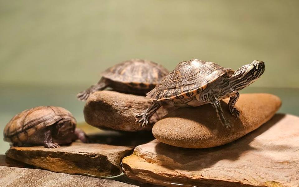
Shell rot is an infectious disease of the keratin scutes and underlying bony shell, triggered primarily by opportunistic bacteria or fungi when husbandry parameters falter. In aquatic species like the African sideneck turtle, moisture-associated shell rot often manifests as soft, discolored patches, pitting, or a pungent odor along the plastron. The microtrauma caused by rough basking docks allows environmental pathogens like Citrobacter or Pseudomonas to penetrate beneath scutes. Left unchecked, the infection can erode through bone directly into the coelomic cavity, turning an external lesion into systemic sepsis.
Terrestrial tortoises, including humid-forest species such as the red-foot tortoise, face a distinct presentation of shell rot. While red-foots require consistently elevated relative humidity to maintain respiratory and epidermal health, waterlogged substrate creates an anaerobic breeding ground. Wet organic substrate presses against the underside of the plastron for hours, degrading the protective outer layer and causing flaky, white or reddish erosions. Differentiating between benign keratin wear and active infectious rot requires close tactile inspection, as true rot softens the bone and bleeds easily when probed gently.
Clinical diagnosis typically involves cytologic evaluation, deep tissue bacterial and fungal cultures, and whole-body radiographs to assess structural invasion. Superficial lesions may respond to gentle surgical debridement, topical antiseptics, and dry-docking protocols tailored strictly to the species. Advanced osteomyelitis requires systemic antibiotics and ongoing therapeutic flushing. Keep in mind that topical treatments applied over necrotic keratin cannot penetrate to the infection base; true healing only begins once dead bone is removed by experienced hands under sterile conditions.
Emergency Triage and Veterinary Repair for Broken Shells
A cracked or crushed shell represents an orthopedic emergency requiring immediate stabilization rather than amateur adhesive fixes. Chelonia shells are living bone covered by keratin, housing the spine, ribs, lungs, and spinal cord immediately beneath the dorsal carapace. When a turtle or tortoise suffers blunt force trauma from a vehicle strike, garden tool, or dog bite, the internal coelomic membrane is frequently compromised. Pathogens enter the bloodstream within hours, making prompt wound lavage and stabilization paramount before any mechanical closure is attempted.
Initial triage focuses on managing pain, combating neurogenic shock, and stabilizing respiratory function. Because chelonians lack a muscular diaphragm and rely on body wall movement to breathe, significant carapace fractures can collapse negative pressure mechanics. A qualified clinician at an experienced reptile veterinary clinic will flush contaminated debris using warmed sterile saline, obtain multi-view radiographs to evaluate organ displacement, and administer multimodal analgesia using appropriate exotic opioid and anti-inflammatory protocols.
Permanent shell repair techniques have evolved past outdated epoxy and fiberglass patches, which frequently trapped anaerobic bacteria and caused fatal underlying osteolytic infections. Modern orthopedic repair employs orthopedic wire, cerclage bridges, zip-tie anchors bonded with non-toxic dental acrylic, or vacuum-assisted closure dressings. These systems stabilize fracture margins while allowing the veterinarian periodic access to flush the healing bed underneath. Depending on the depth of the fracture and patient age, complete bony bridging can take between six months to two full years.
Understanding Shell Pyramiding and Metabolic Bone Imbalances
Shell pyramiding refers to the abnormal vertical growth of individual vertebral and costal scutes, creating raised, conical mounds instead of a smooth, uniform carapace contour. Historically, pyramiding was blamed exclusively on excessive dietary protein, but recent decades of research have proven that microclimatic humidity is the primary developmental factor. In the wild, juvenile tortoises spend their formative years wedged into humid micro-burrows, leaf litter, and deep subterranean crevices, which keeps the emerging keratin layers malleable as bone expands beneath them.
When captive young tortoises are housed under intense heat lamps in open-topped enclosures with dry ambient air, the outer keratin sheath dries out prematurely, stiffens, and exerts mechanical resistance against normal lateral growth planes. The underlying bone continues to expand along the path of least resistance, forcing the scute apex upward into a pyramid. While existing pyramiding is permanent once bone calcifies, correcting the environmental humidity immediately halts progressive deformity in emerging growth rings across juveniles.
Pyramiding is frequently compounded by nutritional metabolic bone disease, where insufficient dietary calcium or lack of effective UVB exposure forces the body to mobilize minerals from skeletal reserves. A tortoise suffering from both conditions will display a pyramid-shaped carapace that paradoxically feels spongy, yielding, or soft under modest finger pressure. Proper therapy requires balanced calcium supplementation with appropriate phosphorus ratios, validated high-output UVB lighting, and the immediate creation of warm, humid microclimates inside the enclosure.
Overgrown Beaks and When a Chelonian Needs a Trim
Tortoises and turtles possess a specialized keratinous structure called the rhamphotheca, which covers the jaws and serves the functional role of teeth. In nature, constant grazing on fibrous grasses, tough weeds, and calcium-dense vegetation naturally wears this keratin sheath down at the same rate it regenerates. In captive settings where soft greens, commercial pellets, or overly diced vegetables predominate, mechanical abrasion fails to occur, leading directly to a rhamphotheca overgrowth that resembles a distorted, sharp beak.
An overgrown beak interferes with the animal's ability to shear foliage, leading to malnourishment, progressive weight loss, and tongue trauma. In severe cases, the upper rhamphotheca can curl inward into the lower jaw or crack vertically up into the vascularized tissue of the maxilla. Attempting to cut an overgrown beak at home using human nail clippers or wire cutters often shatters the brittle keratin along natural grain lines, causing acute bleeding and exposing sensitive underlying bone to opportunistic infections.
Clinical beak trims are performed using high-speed rotary dremel burs or specialized veterinary dental tools while the animal's head is gently restrained. Clinicians contour the biting margins back to anatomical alignment, respecting the underlying vascular germinal matrix. Following a corrective procedure, owners must adjust dietary fiber levels and feed meals on natural rough surfaces, like untreated slate tiles or terracotta saucers, which continuously hone the rhamphotheca during daily feeding.
Normal Shedding Versus Dysecdysis Across Common Geckos
Shedding, or ecdysis, is a complex hormonal and physiological process in which reptiles regenerate their epidermis and slough off the old, worn outer layer of keratin. In common terrestrial species like the leopard gecko, shedding typically occurs every two to four weeks depending on age and growth velocity. Prior to a shed, the gecko's skin appears dull, turns opaque white, and loosens as lymphatic fluid lubricates the plane between old and new tissue layers. Most geckos consume their shed skin to recycle valuable nutrients.
Dysecdysis occurs when the old skin fails to separate cleanly, leaving retained patches across digits, the tail tip, and the ventral scales. On digits, retained rings of dry skin act like microscopic tourniquets, restricting capillary perfusion to peripheral tissue. If left unresolved over successive shed cycles, the starved toe tissue necroses, dries out, and sloughs off permanently. Similarly, retained spectacles over the eyes can cause secondary keratitis, corneal clouding, and progressive visual impairment that disrupts the lizard's predatory strike reflex.
Veterinary intervention for severe dysecdysis involves therapeutic warm-water soaks, application of sterile ophthalmic lubricants, and careful manual debridement under magnification. Owners should never pull dry, adherent skin off a gecko forcefully, as this rips immature living scales beneath and creates open dermal wounds. Providing a permanent humid hide filled with damp sphagnum moss inside the terrarium resolves the overwhelming majority of minor shedding difficulties before veterinary extraction becomes necessary.
Managing Shed Cycles in Arboreal Geckos and Corn Snakes
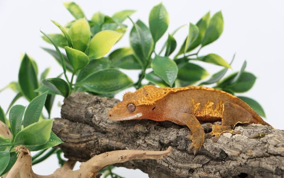
Arboreal species, such as crested geckos, exhibit distinct shedding vulnerabilities tied closely to their specialized lamellae and scansor pads. Crested geckos rely on microscopic hair-like structures, or setae, on their toe pads to adhere to slick vertical surfaces. When dry shed fragments adhere to these pads, the lizard loses traction, leading to falls and chronic stress. Crested geckos do not possess eyelids; instead, they have transparent scales called spectacles that must cleanly release with each shed cycle to avoid blinding build-ups.
In colubrid snakes like the corn snake, a normal shed should detach in one single, continuous, inverted tube from the rostral tip to the tail tip. During the initial 'blue' phase of ecdysis, the snake's eyes turn milky blue-grey and temperament becomes defensive due to compromised vision. If environmental humidity drops below ideal thresholds during this week-long cycle, the skin dries out and tears into dozens of brittle flakes, frequently leaving the ocular spectacles or the very tip of the tail trapped.
If you observe a corn snake struggling through fragmented shedding cycles across multiple months, consult with exotic pet veterinarians to evaluate both enclosure humidity curves and underlying parasitic or systemic infections. Chronic dysecdysis is frequently a clinical symptom of subclinical dehydration, low ambient temperatures, or internal protozoal burdens. Adjusting misting routines, replacing screen enclosure tops with moisture-retentive barriers, and verifying ambient hydration levels usually restores normal full-body shed mechanics.
Digestive Impaction in Terrestrial Lizards
Digestive impaction is the mechanical blockage of a reptile's gastrointestinal tract by indigestible foreign material or hardened fecal masses. In leopard geckos, impaction most frequently involves calcium carbonate sands, walnut shells, loose gravel, or oversized feeder insects with dense chitinous exoskeletons. While wild geckos occasionally ingest small mineral grains, captive geckos suffering from subclinical calcium deficiencies often display geophagy—the deliberate consumption of substrate in an instinctual effort to acquire deficient trace minerals.
A gecko suffering from gastrointestinal obstruction presents with progressive lethargy, abdominal distention, lack of fecal output, and visible anorexia. In light-bellied species, an unnatural dark or bluish mass may be visible through the ventral body wall, representing congested intestines or necrotic loops of bowel. As food rots inside the upper digestive tract, the gecko may regurgitate undigested insects, develop secondary bacterial enteritis, and dehydrate rapidly due to fluid loss into the intestinal lumen.
Clinical treatment depends on the severity and location of the obstruction confirmed via lateral and dorsoventral radiographs. Mild cases often resolve with thermal stabilization, subcutaneous fluid therapy, oral osmotic laxatives, and guided lukewarm shallow baths to stimulate gut peristalsis. Dense, completely obstructive masses or cases involving gut perforation demand emergency surgical celiotomy to exteriorize and clear the affected bowel before fatal peritonitis develops.
Diagnostic Workups in an Exotic Clinic Setting
Exotic veterinary examinations differ markedly from domestic companion animal visits due to the subtle clinical presentation typical of ectotherms. Reptiles are instinctual prey species that mask severe physiological distress until their metabolic reserves are almost entirely depleted. A thorough clinical evaluation begins with a meticulous assessment of the owner's home husbandry parameters, including hot spot temperatures, night drops, thermal gradient ranges, humidity percentages, and the exact age and brand of UVB lamps.
Diagnostic imaging plays a foundational role in identifying internal pathology that cannot be palpated through scales or shell. Digital radiography reveals bone density alterations indicative of metabolic bone disease, outlines radiopaque foreign bodies in the gastrointestinal tract, and visualizes the coelomic extent of shell fractures. For complex shell fractures or deep soft-tissue abscesses, advanced practitioners often use computed tomography, which provides cross-sectional slices free from the diagnostic shadowing created by dense bone.
Hematology and biochemistry profiles offer crucial insights into internal organ function, hydration status, and systemic inflammatory responses. Blood samples collected from the jugular vein, subcarapacial sinus, or ventral coccygeal vein allow clinicians to quantify white blood cell responses, calcium-to-phosphorus ratios, and uric acid concentrations. When residents in high-population regions seek qualified assistance, facilities like exotic pet vets in California or exotic pet vets in Florida frequently provide in-house specialized clinical pathology calibrated specifically for ectotherm blood chemistry.
At-Home Supportive Care and Quarantine Environments
When managing an injured or recovering reptile, the primary captive habitat must be temporarily decommissioned in favor of a sterile hospital enclosure. Standard loose substrates like coconut fiber, cypress mulch, or sand must be completely removed, as particles will adhere to open shell fractures, enter unhealed surgical sites, or get consumed by uncoordinated animals. Unprinted paper towels or plain butcher paper serve as the gold standard for hospital flooring, providing a clean surface that makes monitoring droppings and wound discharge straightforward.
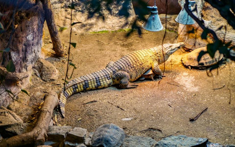
Environmental parameters within a quarantine tank require heightened precision. Recovering reptiles must be kept at the species-specific preferred optimum temperature zone (POTZ) continuously to support cellular repair and drug metabolism. Dropping below this temperature range impairs the reptile's innate immune response and retards the clearance of veterinary antibiotics. Conversely, excessive heat accelerates systemic dehydration, especially in animals receiving osmotic laxatives for impaction or undergoing daily debridement for shell rot.
Soaking protocols should be conducted under strict veterinary supervision and clear guidelines. For patients recovering from shedding disorders or mild impactions, lukewarm shallow soaks should never exceed the bridge height of the plastron or the shoulder height of the lizard. Animals with open shell cracks or active surgical incisions should rarely be submerged in water baths, as bathwater carries opportunistic contaminants directly into deep tissue. Instead, wound-safe dressings and isolated humid hides maintain necessary tissue moisture safely.
Long-Term Prevention Through Environmental Calibration
The overwhelming majority of reptile shell disorders, chronic dysecdysis, and gastrointestinal impactions stem directly from environmental mismanagement rather than innate fragility. Reptiles are entirely dependent on their artificial surroundings to regulate body temperature, synthesize vitamins, and maintain cellular hydration. Establishing an accurate baseline requires ditching analog dial thermometers and hygrometers, which are notoriously inaccurate, in favor of digital probes and infrared non-contact temperature guns to measure real surface temperatures.
Full-spectrum ultraviolet lighting is an indispensable pillar of preventative husbandry that directly prevents metabolic deformities and pyramiding. Linear T5 fluorescent fixtures emit predictable levels of UVB radiation across wide basking zones, whereas compact coil bulbs frequently produce uneven, concentrated hotspots that drop off sharply beyond a few inches. UVB lamps degrade over time and must typically be replaced every six to twelve months, regardless of whether the bulb still visibly illuminates, because the phosphor coating decays before visible light ceases.
Finally, balanced diets matching the evolutionary biology of the species mitigate behavioral deficiencies that drive animals to ingest foreign substrates. Providing dietary variety, dusting feeder insects with high-grade calcium carbonate lacking added vitamin D3 for animals with continuous UVB access, and providing a clean water source daily ensures organ systems operate smoothly. Preventative wellness examinations conducted annually by a reptile specialist catch subtle carapace softening, early beak misalignments, and weight plateaus before emergency hospitalization becomes necessary.
Published 2026-09-21 · reviewed with each rebuild, last September 2026.
Find exotic pet vets near you
The directory lists 3,226 exotic pet vets by state and city — pick your state or use the “Near me” button in the header.
More guides
-
 Owning
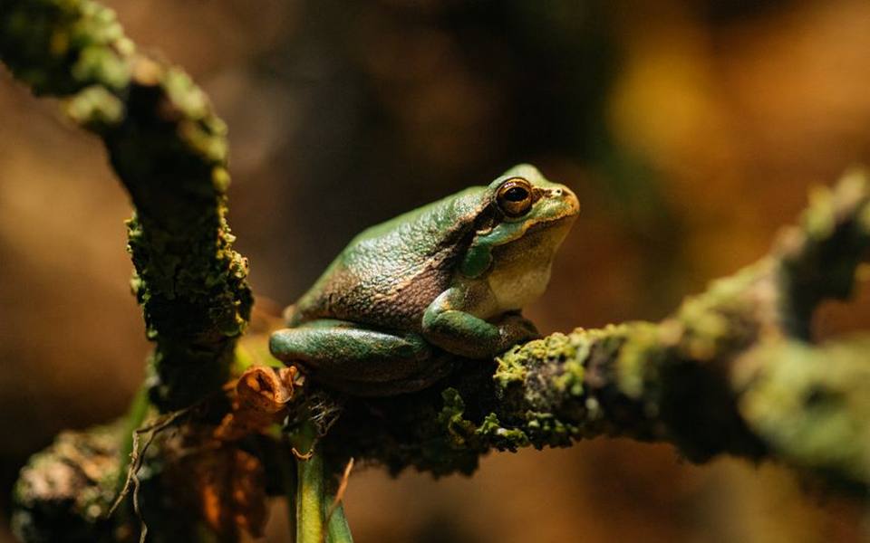
What Are Amphibians? A Complete Guide to Amphibians as Exotic Pets
-
 Money
MoneyHow to Find Affordable and Emergency Vet Care for Exotic Pets
-
 Money
MoneyHow Much Does a Parrot Cost? Complete Exotic Bird and Pet Care Price Guide
-
 Owning
Can You Keep a Capybara as a Pet? Legality, Care, and Diet Requirements
-
 Owning
How to Treat and Get Rid of Snake Mites Safely
-
 Owning
Keeping Exotic Mammals: Can You Have a Fennec Fox or Kinkajou as a Pet?
-
 Money
MoneyLow-Cost Rabbit Neutering: What to Expect and How to Find Affordable Care
-
 Owning
What Is an Avian Pet? A Guide to Bird Care and Specialized Veterinary Medicine
-
 Service
How to Find and Choose the Right Exotic Pet Veterinarian
-
 Owning
Swim Bladder Disease in Aquarium Fish: Causes, Symptoms, and Treatment
-
Owning
How to Identify and Treat Mites and Skin Parasites in Reptiles, Snakes, and Rabbits
-
Safety
Rabbit GI Stasis Guide: Symptoms, Emergency Care, and Prevention
-
 Owning
Bearded Dragon Health Guide: Brumation, Impaction, Fungus, and Skin Issues
-
 Safety
Hamster Wet Tail Disease: Causes, Symptoms, Treatment, and Prevention
-
 Owning
Clinical Care Guide: Common Illnesses in Ferrets, Pythons, and Rabbits
-
Owning
Sugar Glider Diet and Feeding Guide: BML, TPG, and Nutritional Balance
-
Owning
Bird Grooming Guide: Wing Trimming, Beak Care, and Nail Maintenance
-
 Money
MoneyExotic Pet Insurance and Vet Costs: What Is Covered and Is It Worth It?
-
 Owning
Legal Exotic Pets and Ownership Laws: Permits, Licenses, and State Regulations
-
Owning
Bird Feather Plucking and PBFD: Causes, Symptoms, and Diagnostic Steps
-
 Service
Psittacine Health and Avian Veterinary Care Guide
-
 Service
How to Find and Evaluate Exotic Pet Clinics, Specialists, and Emergency Care
-
 Owning
Small Mammal Healthcare: Managing Chronic Illness and Emergencies
-
 Safety
Avian Egg Binding and Yolk Peritonitis: Emergency Care and Clinical Protocols
-
 Owning
Bird Grooming, Wing Maintenance, and Early Illness Signs
-
 Service
Backyard Poultry and Waterfowl Veterinary Care Guide
-
 Buying
Exotic Pet Ownership: Legal Rules, Realistic Costs, and Beginner Options
-
 Service
Aquatic Medicine: Treating Swim Bladder and Fish Infections
-
 Money
MoneyRabbit Spay, Neuter, and Vaccination Costs: Complete Veterinary Guide
-
 Owning
Understanding Amphibians: Characteristics, Classification, and Pet Care Basics
-
 Owning
Bearded Dragon Brumation and Metabolic Bone Disease: Signs, Causes, and Care
-
 Buying
Parrot Ownership Guide: Prices, Types, and Avian Basics
-
 Owning
Complete Guide to Snake Mite Treatment: Products, Safe Sprays, and Vet Care
-
 Money
MoneyRabbit Spay and Neuter Costs: Vaccinations and Low-Cost Vet Options
-
 Owning
What Are Amphibians? Traits, Classification, and Pet Care Basics
-
 Owning
How to Get an Exotic Pet License: Legal Permits and Animal Ownership Laws
-
 Owning
Reptile Health Guide: Bearded Dragon Brumation, Metabolic Bone Disease, and Snake Mite Control
-
 Money
MoneyRabbit Spay, Neuter, and Vaccination Costs: Low-Cost Vet Guide
-
 Money
MoneyHow Much Does an Exotic Vet Cost? Emergency, Routine, and In-Home Care Guide