Recognizing Avian Reproductive Crises
Egg binding and egg yolk peritonitis represent two of the most urgent medical crises seen in captive female birds. When an egg lodges in the reproductive tract or ovum material spills into the abdominal cavity, a hen can deteriorate within hours. The high metabolic rate of companion birds means systemic shock, pressure on vital organs, and fatal sepsis can develop rapidly without prompt intervention. Owners must act swiftly at the first hint of distress rather than waiting to see if the situation resolves on its own.
Both conditions stem from complications within the avian reproductive anatomy, specifically the ovary and the oviduct. In a healthy cycle, an ovulated follicle travels down the infundibulum, magnum, isthmus, and uterus before exiting through the cloaca. When mechanical blockage, physiological exhaustion, or ectopic ovulation disrupts this pathway, severe tissue inflammation and circulatory compromise follow. Understanding that these problems rarely resolve spontaneously is the first step in saving an affected bird's life.
Survival depends almost entirely on how fast the bird receives supportive care and professional medical attention. Whether dealing with a tiny finch or a large cockatoo, reproductive complications put tremendous pressure on the renal system, respiratory air sacs, and caudal blood flow. Stabilizing the bird in a quiet, warm environment while contacting a clinical facility offers the best chance of navigating these high-stakes emergencies successfully.
Understanding the Differences Between Dystocia and Peritonitis
Although owners frequently group these reproductive issues together, egg binding and peritonitis involve distinct pathological processes that require different treatment strategies. True egg binding, clinically termed dystocia, occurs when a formed or partially formed egg becomes stuck inside the caudal reproductive tract, usually within the uterus, vagina, or cloaca. The physical presence of the egg creates mechanical obstruction, compressing the kidneys and sciatic nerves, which can lead to rapid hindlimb paresis, severe metabolic acidosis, and circulatory collapse.
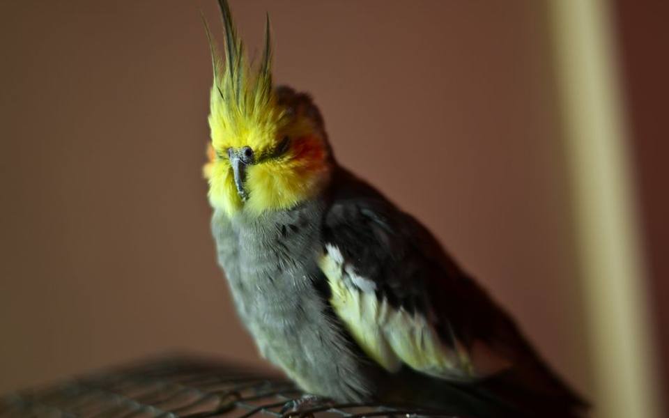
Egg yolk peritonitis, by contrast, happens when liquid yolk material enters the coelomic cavity instead of traveling through the oviduct. This can occur if an ovum ruptures during oviductal contraction, misses the infundibulum during ovulation, or if an impacted egg inside the reproductive tract breaks under pressure. The lipid-rich, protein-dense yolk acts as an intense chemical irritant against the sensitive coelomic serosa, triggering massive non-infectious inflammation that quickly attracts opportunistic bacteria from the gastrointestinal tract.
In many chronic cases, the two disorders develop side by side. A hen that has struggled with an obstructing shell for several days may suffer oviductal rupture, discharging infectious yolk fluid directly into her internal cavities. Alternatively, pre-existing subclinical coelomitis can cause muscle fatigue and scarring along the oviduct walls, preventing normal peristalsis and trapping subsequent eggs. Differentiating between purely mechanical dystocia and diffuse peritonitis often requires specialized imaging by experienced avian care professionals.
Species Vulnerability Across Parrots, Budgies, and Cockatiels
Reproductive emergencies do not distribute evenly across all bird families, with certain companion species showing extreme susceptibility. Cockatiels, budgerigars, lovebirds, and canaries are notorious for chronic, persistent egg production even in the absence of a male partner. Small body sizes and selective breeding for prolific reproduction have left many small parrots with narrow pelvic canals and easily depleted nutritional reserves, amplifying their overall risk of life-threatening binding events.
Cockatiels in particular are considered poster hens for excessive hormonal stimulation and chronic ovulatory cycles. A female cockatiel kept on an unbalanced seed diet will frequently lay multiple clutches throughout the spring and autumn months, gradually stripping calcium carbonate from her skeletal system to supply eggshell synthesis. As circulating calcium plunges, the muscular walls of her uterus lose the contractile force needed for expulsion, stranding the egg just inches from the vent.
While budgies and small parakeets face high rates of dystocia due to their diminutive physical frames, larger parrots are not entirely immune. Amazons, macaws, and African greys develop reproductive complications less frequently, but when they do, the underlying triggers often involve severe oviductal cystic disease, salpingitis, or obesity-induced coelomic crowding. Regardless of the patient's physical size, any hen actively producing eggs should be monitored with intense vigilance throughout her laying cycle.
Visible Signs, Straining, and Dropping Changes
Detecting a reproductive emergency early requires knowing how a sick bird disguises pain and identifying subtle behavioral changes. A hen experiencing dystocia typically presents fluffed on the bottom of her enclosure, unwilling to perch, with her eyes half-closed and wings held slightly outward from her body. You may observe rhythmic tail bobbing, wide-stance perching, and visible abdominal straining as her body attempts to pass the blockage against swollen cloacal tissues.
Changes in droppings offer crucial diagnostic clues for both conditions, especially in cases of suspected yolk peritonitis. When active yolk leakage occurs, bird droppings frequently shift from firm, uniform structures to loose, pale yellow or pea-green puddles. In severe cases, the cloacal discharge takes on an oily, curdled appearance resembling scrambled egg material mixed with chalky urates, signaling that broken yolk proteins are flushing through the lower intestinal tract alongside systemic bile pigments.
Droppings may also cease entirely if a large, bound shell physically compresses the lower intestine and ureters against the pelvic ceiling. Complete absence of fecal output accompanied by persistent straining constitutes a dire emergency requiring immediate veterinary transport. Any sudden swelling around the vent, bluish discoloration of the cloacal mucosal tissue, or weakness in one or both legs indicates severe nerve compression that can become permanent if unaddressed.
Underlying Causes of Avian Reproductive Failure
Reproductive complications rarely occur in a vacuum; they almost always stem from a combination of nutritional deficiencies, environmental mismanagement, and hormonal imbalance. Nutritional secondary hyperparathyroidism, caused by diets dominated by dry seed mixes that lack essential calcium and vitamin D3, remains the leading cause of soft-shelled eggs and uterine muscle fatigue. Without adequate ionized blood calcium, smooth muscle cells simply cannot generate the sustained contractions needed for oviposition.
Environmental triggers also trick a hen's endocrine system into believing she must continually produce offspring. Extended artificial daylight exposure of fourteen hours or more, constant access to high-fat foods, warm baths, and perceived nesting sites such as cardboard boxes or hollow toys all stimulate follicle-stimulating hormone. When these breeding stimuli remain present year-round, the hen's reproductive machinery runs continuously without the dormant recovery phase naturally provided by seasonal shifts.
Physical and pathological oviductal anomalies further exacerbate the situation. Salpingitis, an inflammation or infection of the oviduct, can result from ascending bacterial infections, leaving fibrous scar tissue that constricts the lumen. Similarly, obesity causes expansive fat deposits inside the coelom, physically narrowing the path an egg must travel while placing excessive strain on the liver and circulatory system during labor.
Safe First Aid and Transport Strategies
When an owner suspects an egg is stuck, providing appropriate supportive care before departing for an emergency clinic can mean the difference between survival and death. The single most important environmental parameter to adjust is ambient temperature. Move the hen to a small, warm hospital cage or a padded transport carrier kept between eighty-five and ninety degrees Fahrenheit, as hypothermic birds rapidly exhaust their energy reserves trying to stay warm.

Elevating ambient humidity to roughly sixty or seventy percent helps relax tense muscular tissues around the vent without causing undue stress. A quiet, dimly lit bathroom filled with warm shower steam can serve as a temporary stabilization chamber for ten to fifteen minutes while arranging transport. Do not place the bird directly in hot water or force her to endure extreme heat, which can easily induce fatal thermal stress in an already compromised respiratory state.
Critically, owners must avoid dangerous folk remedies that circulate across unverified pet forums. Never attempt to manually press, squeeze, or manipulate the hen's abdomen, and never insert kitchen oils or petroleum jelly into her vent. Applying mechanical pressure to an egg frequently fractures the brittle shell inside the oviduct, causing sharp fragments to lacerate internal walls and transforming a manageable dystocia into fatal internal hemorrhage and septic shock.
In-Clinic Veterinary Triage and Diagnostics
Upon arrival at an animal hospital, clinical staff will prioritize patient stabilization over extensive diagnostics. If the hen exhibits severe respiratory distress, profound shock, or extreme hypothermia, the clinical team will immediately place her in a heated oxygen chamber before attempting physical handling. Intravenous or subcutaneous fluid administration may be initiated to correct electrolyte imbalances, expand vascular volume, and combat hypovolemic shock before touching the caudal abdomen.
Once stable, the veterinarian will perform gentle palpation and whole-body digital radiographs to confirm the exact location, size, and orientation of the egg. Radiography reveals whether the shell has calcified normally, whether multiple eggs are queued behind one another, and if skeletal osteomyelosclerosis—the calcium store inside long bones—has been depleted. Facilities providing exotic pet medical care rely heavily on these diagnostic images to guide their intervention pathway.
For suspected yolk peritonitis without an intact shell, coelomic ultrasonography and abdominocentesis are the primary tools. Ultrasound imaging readily reveals free fluid pocketing between intestinal loops and hepatic margins, distinguishing between clear transudates and dense, cellular yolk exudate. Withdrawing a small fluid sample using a fine needle allows cytology and bacterial culture, pinpointing whether the peritonitis remains sterile or has evolved into an active, life-threatening bacterial infection.
Clinical Protocols for Delivering the Egg
If radiographs reveal a fully shelled, properly positioned egg near the cloaca and the hen remains hemodynamically stable, clinicians generally begin with conservative medical management. Injectable calcium gluconate is administered to restore intracellular calcium pools, enabling uterine smooth muscle to resume natural contractions. This is often paired with warm fluid therapy, mild sedation to relieve cloacal spasms, and controlled humidified heat inside an incubator.
When an egg fails to pass despite medical support, or when soft-tissue swelling obstructs the pathway, medical teams often turn to percutaneous or transcloacal ovocentesis. Under mild sedation or general anesthesia, a sterile needle is inserted directly into the visible egg pole through the dilated vent or coelomic wall. The veterinarian aspirates the liquid yolk and albumen, causing the shell to collapse inward so it can be extracted in soft, folded fragments without lacerating mucosal tissues.
Should ovocentesis prove impossible due to severe ectopic placement, oviductal torsion, or physical impaction, emergency celiotomy and salpingohysterectomy become mandatory. Performing surgical intervention on an unstable avian patient carries significant risks, but removing the compromised oviduct eliminates the obstruction permanently. Clinicians practicing at dedicated exotic animal hospitals weigh these surgical variables carefully based on the patient's cardiovascular stability and tissue viability.
Treating Coelomic Inflammation and Sepsis
Managing egg yolk peritonitis requires an aggressive, multi-pronged pharmaceutical approach designed to halt systemic shock, neutralize inflammation, and sterilize the coelomic cavity. Because free yolk material triggers profound serosal inflammation and adhesive fibroplasia, systemic non-steroidal anti-inflammatory drugs like meloxicam are prescribed at therapeutic avian dosages to manage pain and blunt progressive organ adhesions.
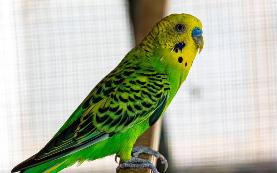
Broad-spectrum antimicrobial coverage is instituted immediately to combat secondary bacterial proliferation, most commonly involving Escherichia coli, Klebsiella, or Enterococcus species translocating across compromised gut barriers. Culture and sensitivity panels derived from coelomic aspirates guide antibiotic selection, ensuring targeted coverage while minimizing toxicity risks to already strained kidneys. In advanced cases, surgical coelomic lavage may be attempted to physically rinse persistent yolk curds from organ surfaces.
Halting ongoing ovarian follicular development is equally vital to keep additional yolk from dropping into an already inflamed coelom. Veterinarians frequently employ hormonal therapy using gonadotropin-releasing hormone agonists, such as deslorelin subcutaneous implants or leuprolide acetate injections. These therapies temporarily shut down the pituitary-gonadal axis, providing a window of reproductive rest during which inflamed tissues can heal without the continuous assault of fresh ovulations.
Long-Term Management and Preventing Relapse
Once an acute crisis has been resolved, long-term survival depends entirely on permanently modifying the hen's environment to dismantle reproductive triggers. Chronic laying must be treated as a behavioral and environmental pathology rather than an inevitable biological reality. Owners must strictly regulate daylight exposure, enforcing a firm regimen of twelve to fourteen hours of uninterrupted, quiet darkness each night to simulate winter conditions that naturally deactivate the gonads.
Dietary overhaul is equally critical to replenishing depleted skeletal stores and stabilizing baseline metabolism. Seed-exclusive regimens must be systematically transitioned to formulated, high-quality extruded pellets supplemented with fresh, dark leafy greens rich in bioavailable calcium, such as kale and dandelion greens. Direct natural sunlight or calibrated full-spectrum avian lighting allows the bird to synthesize endogenous vitamin D3, ensuring efficient calcium absorption and metabolic resilience.
Finally, all social and environmental breeding cues must be eliminated from the living space. Remove mirrors, nesting boxes, fabric huts, shredded paper, and tight dark spaces that mimic nesting hollows. Owners should also adjust how they interact with their pet bird, restricting affectionate petting exclusively to the head and neck while avoiding the back, wings, and cloacal region. Proactively consulting experienced exotic pet veterinary care teams ensures you maintain a safe, non-stimulating routine that keeps reproductive emergencies from returning.
Published 2026-09-21 · reviewed with each rebuild, last September 2026.
Find exotic pet vets near you
The directory lists 3,226 exotic pet vets by state and city — pick your state or use the “Near me” button in the header.
More guides
-
 Owning
What Are Amphibians? A Complete Guide to Amphibians as Exotic Pets
-
 Money
MoneyHow to Find Affordable and Emergency Vet Care for Exotic Pets
-
 Money
MoneyHow Much Does a Parrot Cost? Complete Exotic Bird and Pet Care Price Guide
-
 Owning
Can You Keep a Capybara as a Pet? Legality, Care, and Diet Requirements
-
 Owning
How to Treat and Get Rid of Snake Mites Safely
-
 Owning
Keeping Exotic Mammals: Can You Have a Fennec Fox or Kinkajou as a Pet?
-
 Money
MoneyLow-Cost Rabbit Neutering: What to Expect and How to Find Affordable Care
-
 Owning
What Is an Avian Pet? A Guide to Bird Care and Specialized Veterinary Medicine
-
 Service
How to Find and Choose the Right Exotic Pet Veterinarian
-
 Owning
Swim Bladder Disease in Aquarium Fish: Causes, Symptoms, and Treatment
-
Owning
How to Identify and Treat Mites and Skin Parasites in Reptiles, Snakes, and Rabbits
-
Safety
Rabbit GI Stasis Guide: Symptoms, Emergency Care, and Prevention
-
 Owning
Bearded Dragon Health Guide: Brumation, Impaction, Fungus, and Skin Issues
-
 Safety
Hamster Wet Tail Disease: Causes, Symptoms, Treatment, and Prevention
-
 Owning
Clinical Care Guide: Common Illnesses in Ferrets, Pythons, and Rabbits
-
Owning
Sugar Glider Diet and Feeding Guide: BML, TPG, and Nutritional Balance
-
Owning
Bird Grooming Guide: Wing Trimming, Beak Care, and Nail Maintenance
-
 Money
MoneyExotic Pet Insurance and Vet Costs: What Is Covered and Is It Worth It?
-
 Owning
Legal Exotic Pets and Ownership Laws: Permits, Licenses, and State Regulations
-
Owning
Bird Feather Plucking and PBFD: Causes, Symptoms, and Diagnostic Steps
-
 Service
Psittacine Health and Avian Veterinary Care Guide
-
 Service
How to Find and Evaluate Exotic Pet Clinics, Specialists, and Emergency Care
-
 Owning
Clinical Guide to Reptile Shell Disorders, Shedding Complications, and Impaction
-
 Owning
Small Mammal Healthcare: Managing Chronic Illness and Emergencies
-
 Owning
Bird Grooming, Wing Maintenance, and Early Illness Signs
-
 Service
Backyard Poultry and Waterfowl Veterinary Care Guide
-
 Buying
Exotic Pet Ownership: Legal Rules, Realistic Costs, and Beginner Options
-
 Service
Aquatic Medicine: Treating Swim Bladder and Fish Infections
-
 Money
MoneyRabbit Spay, Neuter, and Vaccination Costs: Complete Veterinary Guide
-
 Owning
Understanding Amphibians: Characteristics, Classification, and Pet Care Basics
-
 Owning
Bearded Dragon Brumation and Metabolic Bone Disease: Signs, Causes, and Care
-
 Buying
Parrot Ownership Guide: Prices, Types, and Avian Basics
-
 Owning
Complete Guide to Snake Mite Treatment: Products, Safe Sprays, and Vet Care
-
 Money
MoneyRabbit Spay and Neuter Costs: Vaccinations and Low-Cost Vet Options
-
 Owning
What Are Amphibians? Traits, Classification, and Pet Care Basics
-
 Owning
How to Get an Exotic Pet License: Legal Permits and Animal Ownership Laws
-
 Owning
Reptile Health Guide: Bearded Dragon Brumation, Metabolic Bone Disease, and Snake Mite Control
-
 Money
MoneyRabbit Spay, Neuter, and Vaccination Costs: Low-Cost Vet Guide
-
 Money
MoneyHow Much Does an Exotic Vet Cost? Emergency, Routine, and In-Home Care Guide