Recognizing Hidden Illness Across Exotic Species
Ferrets, pythons, and rabbits share very little in terms of anatomy or natural history, yet they present a shared challenge to their keepers: an instinct to mask weakness until an illness becomes advanced. In the wild, displaying overt signs of distress makes small mammals and solitary reptiles prime targets for predation. By the time a ball python exhibits wheezing or a rabbit demonstrates a noticeable tilt of the head, the underlying pathology has typically been progressing for days or weeks. Recognizing subtle behavioral changes early remains the most effective way to prevent permanent injury or loss.
Clinical care for these animals requires an understanding of their distinct physiological vulnerabilities. Ferrets possess high metabolic rates and specific immune profiles that demand targeted preventative immunizations, while also rendering them prone to severe vaccine reactions. Ball pythons and other constrictors are ectotherms whose immune defenses rely entirely on thermal gradients, making them particularly vulnerable to superficial necrotic dermatitis and infectious stomatitis when enclosure moisture or temperature fluctuates. Rabbits rely on continuous digestive motility and open-rooted dentition, meaning that secondary stress from an ear infection or respiratory tract colonizer can swiftly trigger systemic collapse.
When health problems arise, scheduling a visit with clinicians who focus on exotic pet medical care is critical. General companion animal protocols rarely translate safely to non-traditional species, and medications commonly tolerated by dogs or cats can be toxic to rabbits or reptiles. Establishing baseline parameters through regular wellness screenings allows you to distinguish normal species-specific eccentricities from emergent clinical symptoms before emergency intervention becomes necessary.
Essential Ferret Vaccinations and Protection Schedules
Preventative medicine for ferrets centers on shielding them against two fatal viral threats: canine distemper virus and rabies. Canine distemper is an airborne pathogen with a mortality rate approaching one hundred percent in unprotected mustelids. Because ferrets are exceptionally susceptible, domestic exposure can occur without direct contact with dogs; contaminated footwear, clothing, or outdoor equipment can easily carry the virus inside your home. A primary series of vaccinations is typically initiated around eight to ten weeks of age, followed by booster doses every three to four weeks until the kit reaches approximately fourteen to sixteen weeks old.
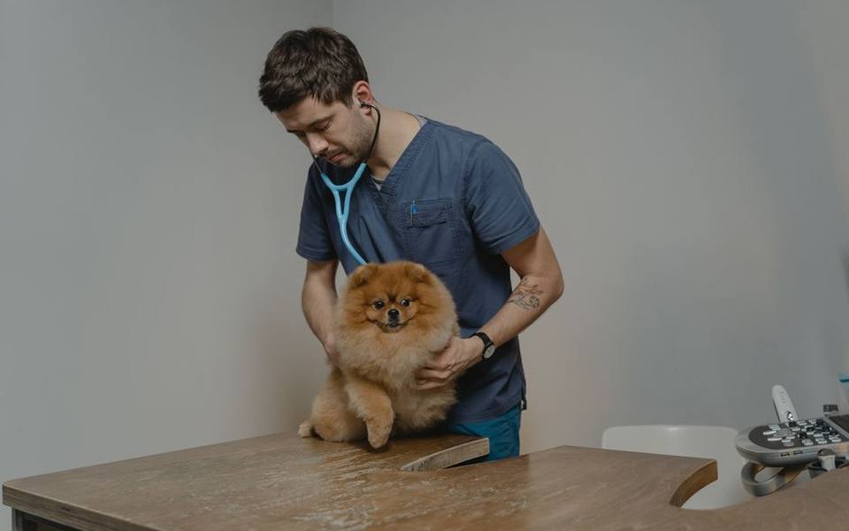
Rabies immunization is both a medical necessity and a legal requirement in most jurisdictions, typically administered as a single dose once the ferret reaches three to four months of age, followed by annual boosters. It is vital to understand that ferrets cannot safely receive standard canine distemper formulations containing modified live virus grown on canine cell lines, as these can actually induce vaccine-induced distemper in mustelids. Only vaccines specifically labeled and approved for ferret use—usually pure recombinant canarypox-vectored vaccines—should be administered by your practitioner.
Routine boosters continue throughout the animal's adult life, but the exact protocol may depend on individual health status, previous reaction history, and regional exposure risks. Keeping strict documentation of every immunization certificate is essential, particularly because local health departments enforce rigid quarantine or testing guidelines for exotic species involved in accidental scratching or biting incidents.
Managing Injection Reactions and Post-Vaccine Care in Ferrets
Ferrets exhibit an unusually high incidence of adverse hypersensitivity reactions following subcutaneous injections, particularly when receiving distemper or rabies biologics. Anaphylactic reactions in mustelids can manifest rapidly, often within fifteen to thirty minutes following the injection. Signs of an acute reaction typically include sudden lethargy, vomiting, pale or cyanotic mucous membranes, profuse watery diarrhea, respiratory distress, and body tremors. In severe cases, circulatory collapse can occur without prompt medical resuscitation.
To minimize these life-threatening risks, experienced practitioners routinely premedicate ferrets with an antihistamine, such as diphenhydramine, roughly twenty to thirty minutes prior to administering the vaccine. Furthermore, gold-standard veterinary care dictates spacing the distemper and rabies injections at least two weeks apart rather than administering them during the same clinical appointment. Splitting the immunizations prevents immune system overload and allows clinicians to pinpoint which agent triggered an adverse event if a reaction does occur.
Following any injection appointment, plan to remain in the veterinary clinic's waiting room for at least thirty to forty-five minutes before traveling home. Once home, monitor the ferret closely for delayed reactions over the subsequent twenty-four hours. Keep the animal warm, provide quiet surroundings, and verify that food and fresh water remain readily accessible. Any signs of facial swelling, hives, breathing changes, or extreme weakness warrant an immediate return to the clinic.
Identifying and Resolving Scale Rot in Pythons
Scale rot, clinically termed ulcerative or necrotic dermatitis, is one of the most common integumentary conditions affecting captive snakes, particularly ball pythons. The disorder begins when a snake rests continuously on damp, soiled, or unhygienic substrate. Excessive moisture softens the lipid layer of the ventral scales, creating microscopic fissures that allow opportunistic environmental bacteria like Pseudomonas, Aeromonas, or fungal spores to invade the dermis. Early signs appear as subtle reddish, yellowish, or brown discolorations along the belly scales.
If left unaddressed, mild discoloration quickly progresses to raised, fluid-filled blisters, shredded scale margins, and open ulcerations that weep serous fluid. This stage is extremely painful for the reptile and introduces a significant risk of systemic septicemia, as bacteria enter the bloodstream through broken cutaneous barriers. Snakes suffering from advanced scale rot often become anorexic, restless, and reluctant to coil normally due to abdominal discomfort.
Treating necrotic dermatitis requires immediate husbandry correction alongside medical intervention guided by qualified reptile care professionals. The animal should be moved to a sterile hospital setup lined with dry paper towels while enclosure humidity is managed through adequate ambient ventilation rather than soaking the bedding. Mild cases may respond to gentle antiseptic soaks, such as heavily diluted povidone-iodine solutions, but deep ulcerations demand diagnostic culture, topical antimicrobial creams, and systemic injectable antibiotics.
Snake Mouth Rot and Infectious Stomatitis
Infectious stomatitis, widely known among reptile hobbyists as mouth rot, represents a progressive bacterial or fungal infection of the oral tissues. In ball pythons, the disease is rarely a primary illness; instead, it almost invariably develops secondary to chronic physiological stress, suboptimal enclosure temperatures, nutritional deficiencies, or minor oral trauma sustained from substrate ingestion or striking cage decor. When the snake's immune response drops, standard commensal oral flora proliferates out of control.
The earliest indicators of mouth rot include subtle petechial hemorrhages—tiny pinpoint red spots—along the gingival margins, increased salivation, and frequent rubbing of the snout against cage surfaces. As the infection takes hold, thick, pale yellowish or white exudate resembling cottage cheese accumulates within the oral cavity. This caseous debris can deform the jawline, prevent complete closure of the mouth, loosen teeth, and eventually invade the underlying mandibular or maxillary bone tissue.
Treatment requires professional intervention to debride necrotic tissue and clear the airway of obstructive exudate. The veterinary team will typically obtain an oral swab for bacterial culture and sensitivity testing to prescribe targeted antibiotics, avoiding indiscriminate antimicrobial use that can foster resistant pathogens. Throughout recovery, keepers must meticulously monitor ambient temperatures within the enclosure, as reptiles require appropriate thermal support at the upper end of their species range to activate their internal immune mechanisms.
Respiratory Infections in Captive Snakes
Respiratory infections in ball pythons and other constrictors are serious, potentially fatal conditions driven by improper environmental parameters. Snakes lack a diaphragm, which means they cannot cough to clear accumulated mucus from their lungs; they rely entirely on body posturing and thoracic contractions. When ambient humidity drops too low or enclosure temperatures fall below the species threshold, the delicate mucosal lining of the trachea and single functional lung dries out, granting environmental microbes an open pathway.
Symptoms of a lower or upper respiratory infection include audible wheezing, clicking sounds during respiration, open-mouth breathing, and excess bubbling mucus visible at the nostrils or inside the oral cavity. An affected snake will often hold its head vertically elevated against the walls of the enclosure in an instinctive effort to keep its airway patent. Lethargy, refusal of prey items, and defensive behavior born of respiratory distress are common systemic manifestations.
Resolving a snake respiratory infection requires strict environmental adjustments alongside veterinary therapy. Enclosure temperatures must be measured using digital probes to verify that the warm basking spot and ambient cool zones align with clinical requirements. Medical treatment typically combines systemic injectable antibiotics with nebulization therapy utilizing saline and antimicrobial solutions to penetrate deep into the snake's simple saccular lung structures.
Untangling Head Tilt and Neurological Signs in Rabbits
Few medical emergencies cause as much immediate alarm for rabbit owners as the sudden onset of a head tilt, clinically termed torticollis. A bunny presenting with a head tilted sharply to one side, loss of balance, rapid involuntary eye movements known as nystagmus, or uncontrolled rolling across the enclosure is experiencing severe vestibular disruption. This alarming presentation is not an illness in itself, but a symptom of a profound disturbance in the inner ear or the central nervous system.
The two most common clinical culprits behind rabbit head tilt are bacterial otitis interna—deep inner ear infection, often linked to organisms like Pasteurella multocida—and infection by the microsporidian parasite Encephalitozoon cuniculi. Differentiating between these causes requires specialized diagnostic work, including skull radiographs or CT scans to examine the tympanic bullae, paired with serological blood panels measuring E. cuniculi antibody titers. Without imaging and lab work, distinguishing an inner ear abscess from neurological protozoal damage is virtually impossible.
Supportive care forms the cornerstone of rehabilitation while specific pharmaceutical agents take effect. Rabbits afflicted with torticollis often struggle to orient themselves, making eating and drinking hazardous or impossible without assistance. Padded, confined enclosures are vital to prevent trauma from rolling, and veterinary protocols typically incorporate anti-nausea medications, anti-inflammatories, fluid therapy, and assisted syringe feeding to safeguard gastrointestinal health.
Upper Respiratory Disease and Snuffles in Rabbits
The colloquial term 'snuffles' refers to chronic upper respiratory tract disease in rabbits, most frequently sparked by the bacterium Pasteurella multocida, though Bordetella bronchiseptica and Staphylococcal species can also play primary or secondary roles. The infection targets the nasal passages, sinuses, and conjunctival sacs. Rabbits are obligate nasal breathers, meaning any structural inflammation or obstructive discharge within the nasal cavity causes significant respiratory compromise and extreme distress.
Clinical presentations range from mild, intermittent sneezing to severe purulent discharge draining from the nares and eyes. Because rabbits groom their faces meticulously using their front paws, keepers often fail to notice early nasal fluid. Instead, the first unmistakable clue is frequently wet, crusty, or heavily matted fur along the inside of the front forearms. If left unmanaged, snuffles can progress downward into chronic pneumonia or track upward into the tear ducts and inner ear structures.
Managing snuffles demands professional oversight from small-mammal care clinicians. Antibiotic therapy must be selected with extreme caution, as oral administration of certain common antibiotics like penicillins, ampicillin, or lincosamides can wipe out benign cecal microflora, precipitating fatal enterotoxemia. Long courses of rabbit-safe antimicrobials, combined with therapeutic nasal flushes and environmental dust reduction, are commonly required to achieve clinical remission.
Rabbit Dental Anatomy and Preventive Care
Dental pathology represents one of the most widespread health issues encountered in domestic rabbits, largely because of their unique anatomy. Rabbits are hypsodont and elodont, meaning all twenty-eight of their teeth grow continuously throughout their natural lifespan. In a healthy animal, the coarse mechanical abrasion produced by grinding long-strand fibrous vegetation naturally wears the crowns down at the same rate they erupt from the gingiva. When this balance is disrupted, structural complications develop rapidly.
If a rabbit consumes an improper diet low in coarse hay or suffers from congenital malocclusion, the teeth do not wear evenly. This leads to the formation of razor-sharp enamel points, known as spurs, along the tongue side of the lower molars and the cheek side of the upper molars. These spikes lacerate the soft oral mucosa and tongue, creating painful ulcerations. Warning signs include dropping food fragments, selective eating of soft pellets over hay, decreased fecal production, facial swelling, wet chins from chronic drooling, and sudden refusal to eat.
Preventive dental maintenance centers entirely on nutrition: clean grass hay, such as timothy or orchard grass, should make up roughly eighty percent or more of a rabbit's daily dietary intake. Regular oral examinations using specialized illuminated speculums allow veterinarians to detect spurring before deep lacerations occur. Once sharp spurs or root elongations develop, corrective dental trims under general anesthesia are necessary to restore balanced occlusal surfaces.
Navigating Diagnostic Visits and Specialized Treatment
Seeking medical treatment for non-traditional species requires forethought and preparation. When transporting a debilitated reptile, ferret, or rabbit to a medical appointment, preserving physical stability during the trip is paramount. Ferrets and rabbits should travel in secure, hard-sided carriers lined with familiar bedding or absorbent towels; avoid wire-bottom carriers that can catch fragile claws. For pythons, transport in a breathable fabric bag or ventilated plastic tub secured inside an insulated transport box with an external, monitored heat source prevents physiological chilling.
During clinical assessments, detailed historical records prove just as valuable as hands-on physical exams. Keepers should bring documented enclosure temperature logs, humidity records, accurate dietary breakdowns, and recent stool or shed samples if requested. Having exact numbers regarding basking spots and ambient gradients allows the veterinary team to differentiate primary infectious agents from environmental husbandry failures in reptiles immediately.
Whether you are locating care in major metropolitan areas or navigating localized listings such as veterinary practices across California, finding an accredited exotic specialist ahead of an acute crisis remains the single best investment in your animal's longevity. Establishing a working relationship with an experienced exotic clinician guarantees access to species-safe diagnostics, tailored anesthetics, and effective emergency protocols when serious symptoms emerge.
Published 2026-09-21 · reviewed with each rebuild, last September 2026.
Find exotic pet vets near you
The directory lists 3,226 exotic pet vets by state and city — pick your state or use the “Near me” button in the header.
More guides
-
 Owning
What Are Amphibians? A Complete Guide to Amphibians as Exotic Pets
-
 Money
MoneyHow to Find Affordable and Emergency Vet Care for Exotic Pets
-
 Money
MoneyHow Much Does a Parrot Cost? Complete Exotic Bird and Pet Care Price Guide
-
 Owning
Can You Keep a Capybara as a Pet? Legality, Care, and Diet Requirements
-
 Owning
How to Treat and Get Rid of Snake Mites Safely
-
 Owning
Keeping Exotic Mammals: Can You Have a Fennec Fox or Kinkajou as a Pet?
-
 Money
MoneyLow-Cost Rabbit Neutering: What to Expect and How to Find Affordable Care
-
 Owning
What Is an Avian Pet? A Guide to Bird Care and Specialized Veterinary Medicine
-
 Service
How to Find and Choose the Right Exotic Pet Veterinarian
-
 Owning
Swim Bladder Disease in Aquarium Fish: Causes, Symptoms, and Treatment
-
Owning
How to Identify and Treat Mites and Skin Parasites in Reptiles, Snakes, and Rabbits
-
Safety
Rabbit GI Stasis Guide: Symptoms, Emergency Care, and Prevention
-
 Owning
Bearded Dragon Health Guide: Brumation, Impaction, Fungus, and Skin Issues
-
 Safety
Hamster Wet Tail Disease: Causes, Symptoms, Treatment, and Prevention
-
Owning
Sugar Glider Diet and Feeding Guide: BML, TPG, and Nutritional Balance
-
Owning
Bird Grooming Guide: Wing Trimming, Beak Care, and Nail Maintenance
-
 Money
MoneyExotic Pet Insurance and Vet Costs: What Is Covered and Is It Worth It?
-
 Owning
Legal Exotic Pets and Ownership Laws: Permits, Licenses, and State Regulations
-
Owning
Bird Feather Plucking and PBFD: Causes, Symptoms, and Diagnostic Steps
-
 Service
Psittacine Health and Avian Veterinary Care Guide
-
 Service
How to Find and Evaluate Exotic Pet Clinics, Specialists, and Emergency Care
-
 Owning
Clinical Guide to Reptile Shell Disorders, Shedding Complications, and Impaction
-
 Owning
Small Mammal Healthcare: Managing Chronic Illness and Emergencies
-
 Safety
Avian Egg Binding and Yolk Peritonitis: Emergency Care and Clinical Protocols
-
 Owning
Bird Grooming, Wing Maintenance, and Early Illness Signs
-
 Service
Backyard Poultry and Waterfowl Veterinary Care Guide
-
 Buying
Exotic Pet Ownership: Legal Rules, Realistic Costs, and Beginner Options
-
 Service
Aquatic Medicine: Treating Swim Bladder and Fish Infections
-
 Money
MoneyRabbit Spay, Neuter, and Vaccination Costs: Complete Veterinary Guide
-
 Owning
Understanding Amphibians: Characteristics, Classification, and Pet Care Basics
-
 Owning
Bearded Dragon Brumation and Metabolic Bone Disease: Signs, Causes, and Care
-
 Buying
Parrot Ownership Guide: Prices, Types, and Avian Basics
-
 Owning
Complete Guide to Snake Mite Treatment: Products, Safe Sprays, and Vet Care
-
 Money
MoneyRabbit Spay and Neuter Costs: Vaccinations and Low-Cost Vet Options
-
 Owning
What Are Amphibians? Traits, Classification, and Pet Care Basics
-
 Owning
How to Get an Exotic Pet License: Legal Permits and Animal Ownership Laws
-
 Owning
Reptile Health Guide: Bearded Dragon Brumation, Metabolic Bone Disease, and Snake Mite Control
-
 Money
MoneyRabbit Spay, Neuter, and Vaccination Costs: Low-Cost Vet Guide
-
 Money
MoneyHow Much Does an Exotic Vet Cost? Emergency, Routine, and In-Home Care Guide